Bilateral Diaphragmatic Hernia By Fetal Sonography

Congenital Diaphragmatic Hernia Radiology Reference Article

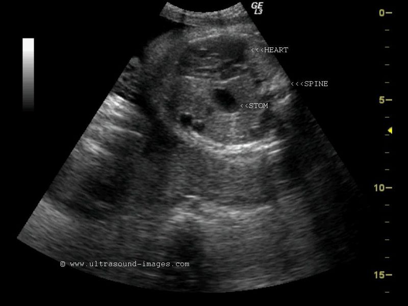
Prenatal Ultrasound At 25 Weeks Of Gestation Shows Right
Bochdalek Hernia

Institute For Advanced Medical Education

Ultrasound And Mr Images Of Prenatally Diagnosed Bilateral

Institute For Advanced Medical Education

C cdh occurs in one of 2000 3000 births.
Bilateral diaphragmatic hernia by fetal sonography. They are primarily pathologies of neonates and most commonly occur unilaterally. Bilateral congenital diaphragmatic hernia was suggested and confirmed by magnetic resonance imaging. In the johns hopkins all children s center for congenital diaphragmatic hernia cdh families find hope during a time that may otherwise feel hopeless. There have been reports of bochdalek hernias in.
The most common is fryns syndrome autosomal recessive. The contents of the hernia range from fat to intra abdominal organs. Detailed ultrasound examination including echocardiography. An autopsy revealed large.
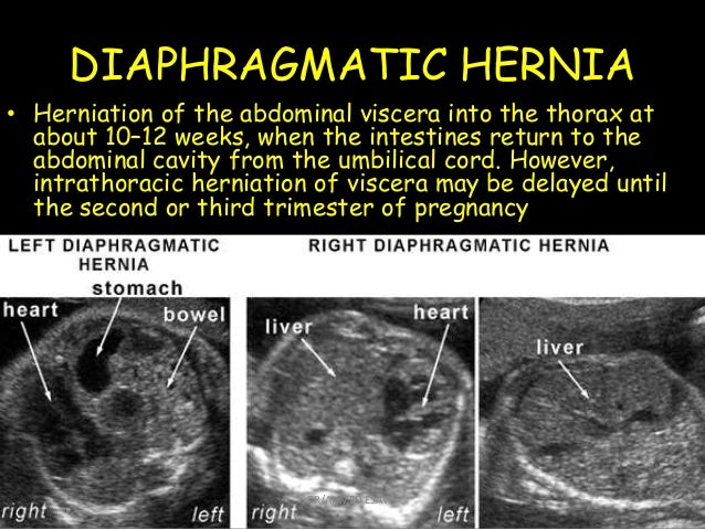
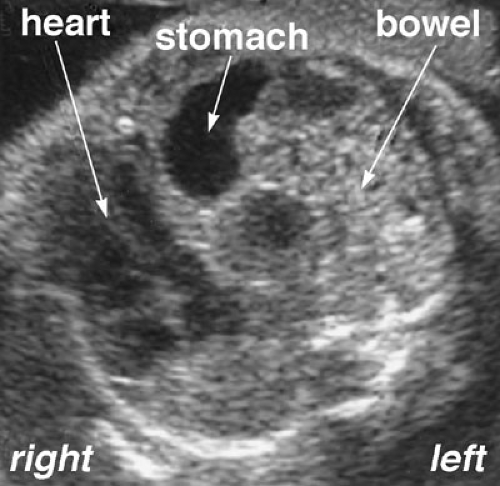
Genetic syndromes are found in 10 of cases. A female newborn weighing 2900 g was delivered at 37 weeks gestation and she died at 7 h of age. A about 97 of cdh are unilateral 1 bilateral and 2 are central hernias. Defects in other systems mainly craniofacial and cardiac are found in 20 of cases.
A bochdalek hernia is a posterolateral diaphragmatic defect that is either congenital or acquired. A baby was delivered at 37 weeks and immediately placed on high frequency oscillation ventilation. These hernias have been described in isolation and as one part of a group of malformations. A defect in the diaphragm of the fetus allows one or more of their abdominal.
Congenital diaphragmatic hernia cdh is a developmental defect of the diaphragm that allows abdominal viscera to herniate into the thorax resulting in pulmonary hypoplasia. Led by cdh expert david kays m d our team combines compassionate care innovative techniques and cutting edge technology and equipment to treat babies with cdh. The presumptive diagnosis was bilateral congenital diaphragmatic hernia.

Prenatal Diagnosis And Management For Congenital Intrapericardial
Prenatal Ultrasound Of Case 2 At 34 Gestational Weeks Shows A A
8 Fetal Chest Dr Ahmed Esawy

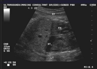
Right Diaphragmatic Hernia
Congenital Diaphragmatic Hernia Obgyn Key

Institute For Advanced Medical Education
A Gallery Of High Resolution Ultrasound Color Doppler 3d

Pulmonary Abnormalities Diagnosis Of Congenital Abnormalities
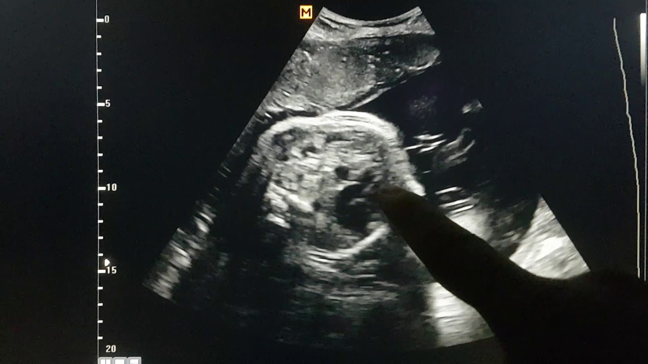
Fetal Diaphragmatic Hernia Sonography Youtube

Prenatal Diagnosis And Perinatal Management Of A Bilateral

Congenital Diaphragmatic Hernia Intechopen

A Review Of Congenital Diaphragmatic Hernia Marlow 2013

Congenital Diaphragmatic Hernia Cdh And Fetal Hydronephrosis
