Uterine Cancer Abnormal Uterus Ultrasound Images

Endometrial Carcinoma In Premenopausal Woman Radiology Case

Ultrasound Characteristics Of Endometrial Cancer As Defined By
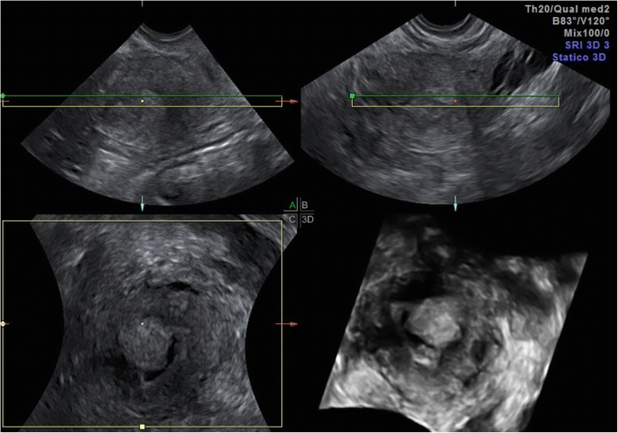
Diagnosing Endometrial Cancer Use 3d Ultrasound First Empowered

Uncovering Endometriosis Vs Endometrial Cancer Empowered

42 Year Old Woman With Abnormal Uterine Bleeding Mdedge Obgyn

Abnormal Uterine Bleeding Houston Causes Of Abnormal Bleeding

For a transvaginal ultrasound a probe that gives off sound waves is put into the vagina.
Uterine cancer abnormal uterus ultrasound images. It can give a perfect insight in the thickness of the endometrium and determine whether there is any hyperplasia i e. For this test the tvus probe that works the same way as the ultrasound transducer is put into the vagina. If you have uterine cancer your. Unlike x rays ultrasound exams do not use radiation.
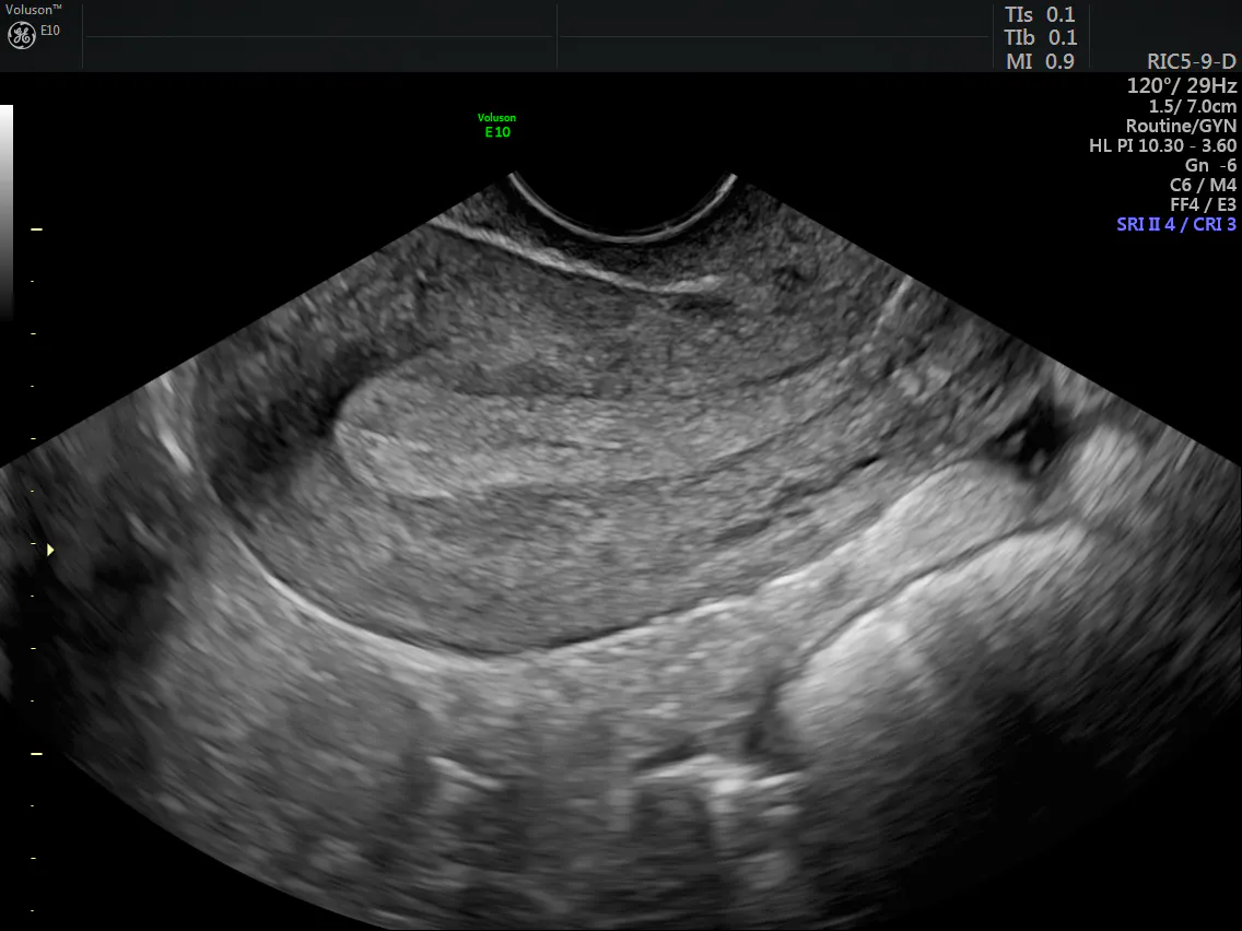
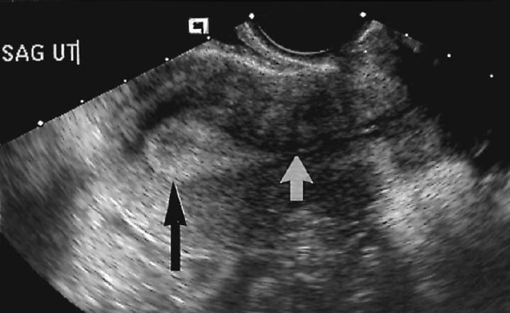
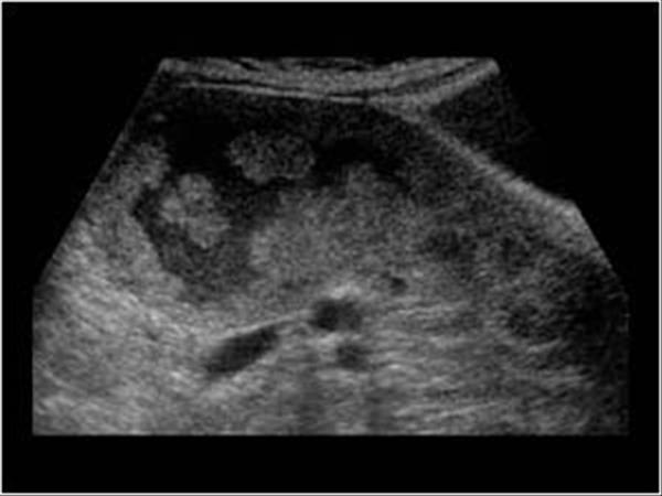
The above ultrasound images show 1 apparently marked thickening of the endometrium 19mm on transabdominal sonography. The sound that tumors produce is different than healthy tissues which helps us identify a uterine tumor. Both ovaries are visible not always the case. By capturing images in real time ultrasound exams reveal the structure and movement of organs such as the heart blood vessels kidneys and liver.
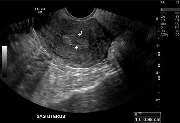
Currently testing for endometrial cancer in women experiencing abnormal vaginal bleeding consists of transvaginal ultrasound imaging biopsy or both. No effective screening method currently exists for women who may be at risk for endometrial cancer. Normal tv image anteverted sagittal. Your doctor may send a thin lighted tube in through your vagina to get a closer look.
Endometrial hyperplasia diagnose with ultrasound ultrasound is a powerful tool used in the diagnosis of many various gynecological conditions. Transvaginal technique anteverted uterus. The overall uterine length is evaluated in the long axis from the fundus to the cervix external. Although people tend to think of biopsies as invasive and frightening an endometrial biopsy is a simple procedure similar to a pap smear dr.
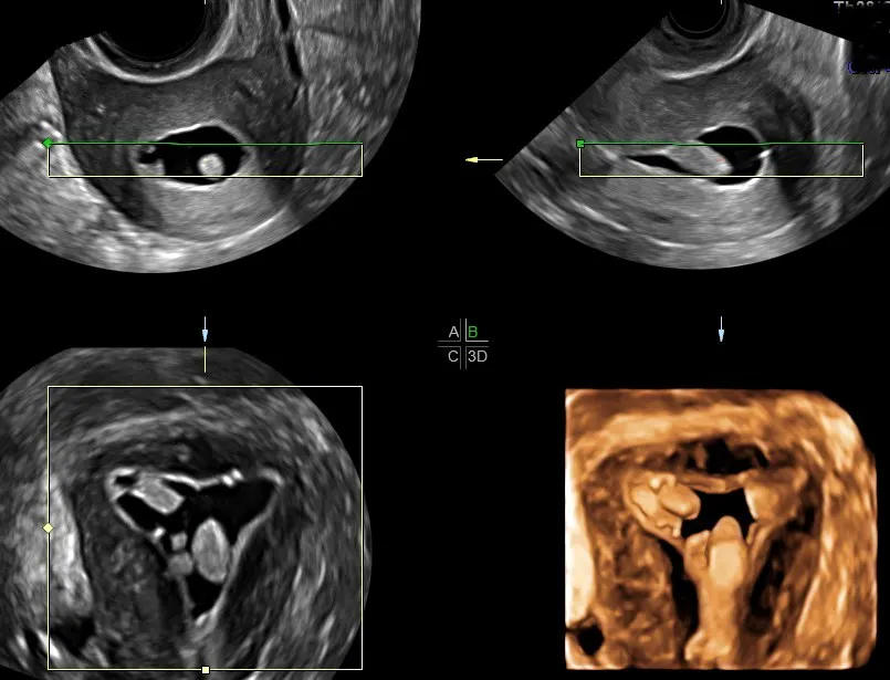
3 color doppler imaging shows feeding vessels supplying the polyp. Trans abdominal view of the uterus. These images can often show if there s a tumor and if it affects the myometrium muscular layer of the uterus. You may have an ultrasound so your doctor can see inside your uterus.
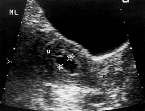
Whether the wall of the uterus is thicker than it is supposed to be. 2 on transvaginal imaging there is a large polyp like mass 14 x 22 mm occupying the uterine cavity. Images from the tvus can be used to see if the uterus contains a mass tumor or if the endometrium is thicker than usual which can be a sign of endometrial cancer. Ultrasound images of malignant polyp of uterus.
The sound waves are used to create images of the uterus and other pelvic organs. This allows physicians to get a better view of the uterus as well as the ovaries. A diagnostic aid such as ultrasound placed in the vagina or transvaginally as opposed to the abdomen can help doctors discover the nature of symptoms. A transvaginal ultrasound tvus is often better to look at the uterus.
Uterine sarcoma is a rare type that starts in the muscles in the uterus or the surrounding tissue. A transvaginal ultrasound produces images of the internal organs when the ultrasound probe is placed in the vagina.

Endometrial Hyperplasia Radiology Reference Article
Endometrial Cancer An Overview Of Novelties In Treatment And

Ultrasound Of Uterus With Cancer Cancer News Update

42 Year Old Woman With Abnormal Uterine Bleeding Mdedge Obgyn

Doctors Seeing More Pcos Women With Endometrial Cancer
Imaging In Endometrial Carcinoma Faria Sc Sagebiel T
Diagnosing Endometrial Cancer Use 3d Ultrasound First Empowered

Abnormal Uterine Bleeding Houston Causes Of Abnormal Bleeding
Endometrial Cancer
Endometrial Polyp With Feeding Vessel Radiology Case
Gynaecology 3 1 Uterus Case 3 1 3 Malignant Uterine And

Transvaginal Ultrasound Showing Measurement Of Tumor
Radiology In The Diagnosis Staging And Management Of Gynecologic
