Bilateral Ventriculomegaly 15mm
Ventriculomegaly

Ventriculomegaly Wikipedia
Congenital Lateral Ventriculomegaly

Fetal Ventriculomegaly Lurie Children S

Ventriculomegaly Radiology Key
The Adventures Of Jnet In Cleveland Update On Babies
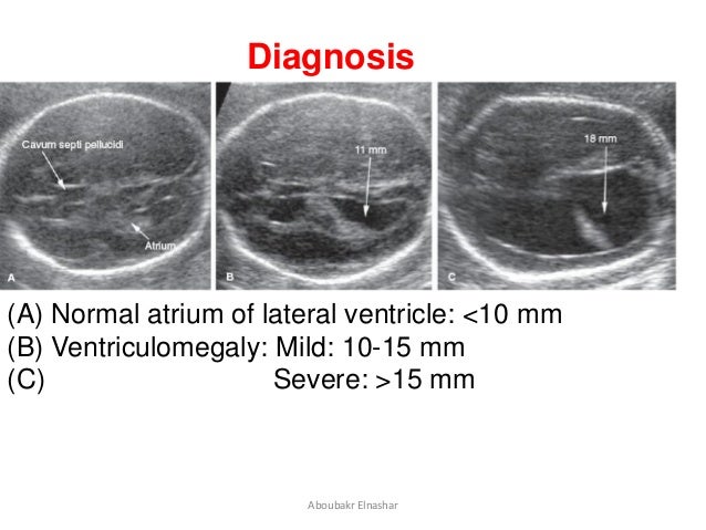
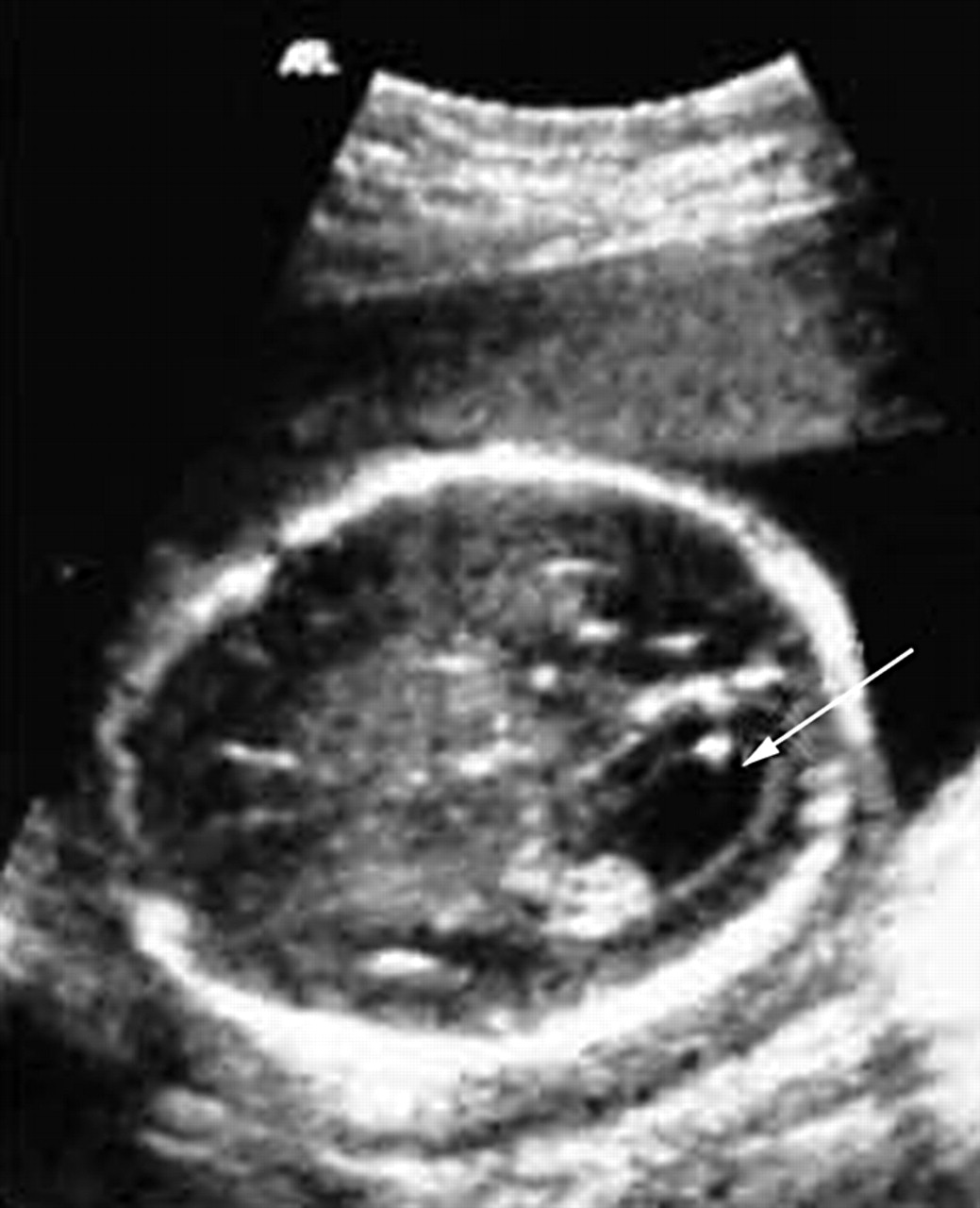
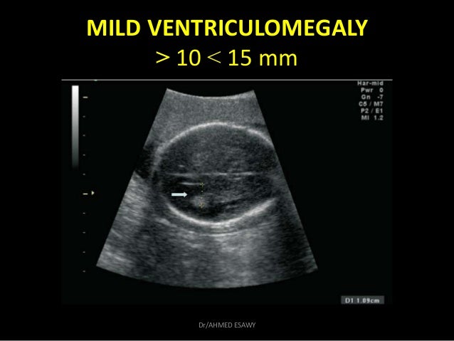
The most common definition uses a width of the atrium of the lateral ventricle of greater than 10 mm.
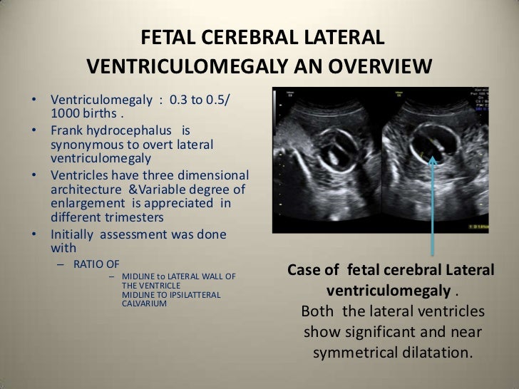
Bilateral ventriculomegaly 15mm. Bilateral or unilateral dilation of the lateral cerebral ventricles observed in the standard transverse section of the brain. This occurs in around 1 of pregnancies. Worsened to 20mm following week. The best outcome is typically observed when.
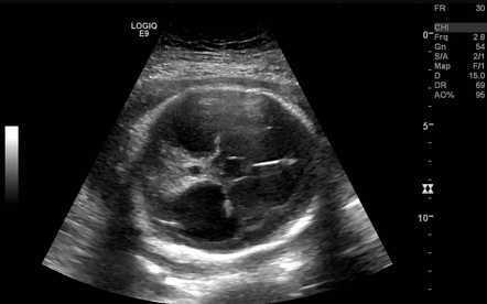
Amino and infection screen clear. Hydrocephalus is the main concern associated with ventriculomegaly. It may occur due to obstruction of cerebrospinal fluid csf flow as a consequence of abnormal development of the ventricles or as part of a destructive process as seen in cerebral atrophy. Ventriculomegaly is a term that describes the actual image of the enlarged spaces as it appears on a prenatal ultrasound.
When the axial diameter measured across the atrium of the ventricle at any gestational age ga exceeds 15 mm ventriculomegaly is said to be severe. If your child has mildly enlarged brain ventricles or ventriculomegaly without other complications the condition may resolve on its own. Ventriculomegaly represents enlargement of the fluid collecting system in the brain. 1 the fetus ventricles are only mildly enlarged measure between 10 15 millimeters in size 2 when there are no other problems seen on the ultrasound and 3 the genetic testing results are normal this is called isolated mild ventriculomegaly.
Hydrocephalus occurs when csf builds up within the ventricles of the brain causing. Hi sorry to hear you are going through this we had bilateral ventriculomegaly at 20 week scan also 17mm each side. When this measurement is between 10 and 15 mm the ventriculomegaly may be described as mild to moderate. This is because excess csf buildup can cause pressure on the brain and neurological problems.
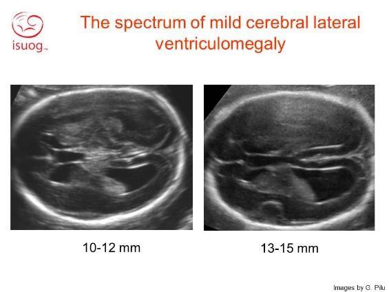
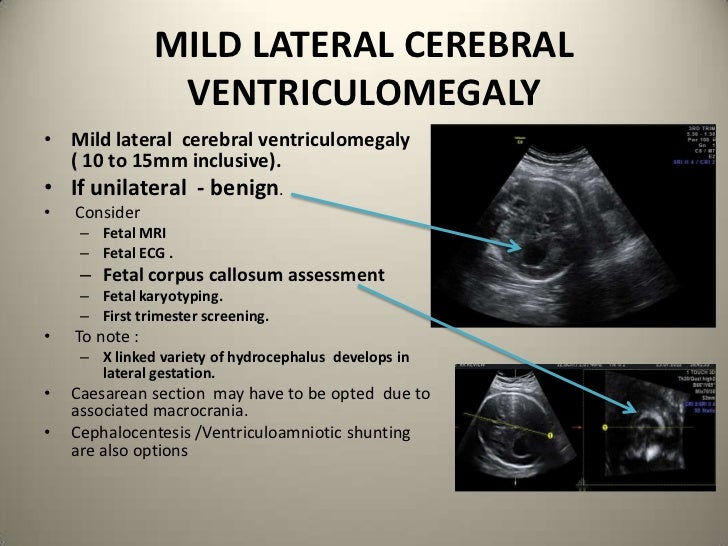
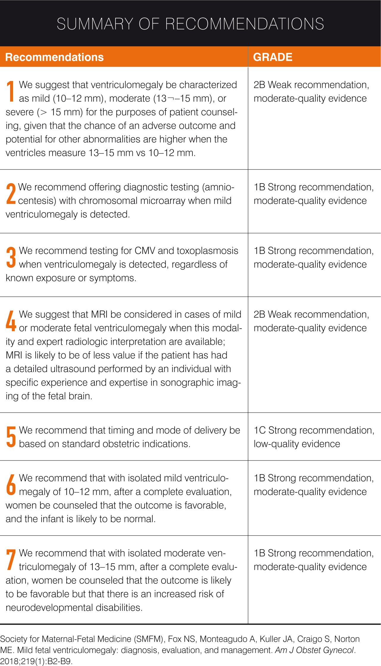
1 5 it is generally agreed that 10 mm is the upper limit of the normal range for the lateral ventricles in an axial plane. Subdivided according to the diameter of the lateral ventricle into mild 10 12 mm moderate 13 15 mm and severe 15 mm. Hydrocephalus is the term used when enlargement of the ventricles has been caused by an increase in the pressure of the cerebro spinal fluid csf within them. When hydrocephalus is more severe or progresses timely treatment is important.
1 5 therefore sonographically diagnosed isolated mild ventriculomegaly imv of the fetus usually refers to cases in which the ventricle sizes are between 10 and 15 mm. This information sheet from great ormond street hospital gosh explains the causes symptoms and treatment of ventriculomegaly and. Enlargement of the ventricles may occur. Are there any medical complications associated with ventriculomegaly.
When the measurement is greater than 15mm the ventriculomegaly may be classified as more severe. Ventriculomegaly is the medical term used to describe enlargement of the ventricles of the brain. Ventriculomegaly is a brain condition that mainly occurs in the fetus when the lateral ventricles become dilated.
Ventriculomegaly Ucsf Fetal Treatment Center
Isolated Mild Fetal Ventriculomegaly Adc Fetal Neonatal Edition
Seven Recommendations For Mild Fetal Ventriculomegaly

Fetal Mild Ventriculomegaly Still A Challenging Problem In

Mild Fetal Ventriculomegaly Diagnosis Evaluation And Management
Https Mydoctor Kaiserpermanente Org Ncal Images Gen Us 20ventriculomegaly 20handout 206 09 Tcm63 18471 Pdf
Fetal Ventriculomegaly Radiology Reference Article Radiopaedia Org
Congenital Lateral Ventriculomegaly

Isolated Mild Fetal Ventriculomegaly Adc Fetal Neonatal Edition
1 Fetal Genetic Ultrasound Dr Ahmed Esawy
Prenatal Genetic Considerations In Congenital Ventriculomegaly And
Congenital Lateral Ventriculomegaly
